Fundus autofluorescence, or FAF is an imaging technique utilized to aid in the documentation and monitorization of retina health and function. The retina is the neurosensory layer of the eye that transmits the optical images we see into the electrical images our brain understands.
The retina contains millions of photoreceptor cells that enable our eyes to see light and color.
There are two types of photoreceptor cells; Rods and Cones
There are approximately 120 million rod photoreceptor cells in the retina. The rods are more sensitive to light, although they are not sensitive to color. The rods aid in peripheral vision as well as in darker environments like at night or when stepping into a movie theatre.
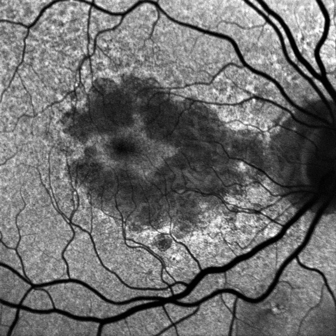

Fundus auto-fluorescent images of the right and left eye exhibiting areas of hypo-fluorescence sparing the central fovea in dry macular degeneration or DRY AMD. In this case, the central vision is mainly spared.
There are approximately 6-7 million cone photoreceptor cells in the retina. The cones are sensitive to color and are most concentrated in the macula where our central, sharpest vision is located. There are three types of cones and they are most sensitive to red, green, and blue light.
Photoreceptor rod and cone cells convert light into the electrical signals transmitted to the brain. They play a key role in our sight. The photoreceptor cells react much like a light bulb; when light reaches them, they fire and then shed their outermost layer in the process.
The retinal pigment epithelium or RPE layer, lies just beneath the photoreceptor cells and is responsible for ingesting the outer segments of the photoreceptor cells in a process called phagocytosis. The molecules may form lipofuscin; a pigmented cell granule that may collect as a part of the normal aging process.
Sometimes, too much lipofuscin may occur causing damage to the retinal pigment epithelium. This excess of lipofuscin may cause degenerative disease of the retina which may need treatment to halt the degeneration. Dry AMD and diseases such as Plaquenil toxicity may advance the process of lipofuscin activity. This FAF photographic technique allows for current assessment and monitorization of progression of RPE disorders. It only takes moments and is completed in the office.
What can I expect during a Fundus Autofluorescent (FAF) Image?
An autofluorescent or FAF image is performed in the office and only takes moments to acquire. The FAF image is often taken alongside other imaging techniques. After a patient is dilated, he/she will be positioned in the chin rest in front of the camera. A safe, bright light is used to fluoresce the lipofuscin in the retinal pigment epithelial layer. Although the light used is bright, it is safe and its peak emission is about 630 nm (nanometers). The light wave utilized enables the physician to evaluate and monitor the stress of the photoreceptor cells. The physician is able to measure areas of atrophy as well as areas of possible degenerative progression. Since the image uses such a bright, safe light, an after-effect color hue may be experienced. This is similar to seeing a bright box in your vision after a family photograph. It is normal and will subside momentarily.